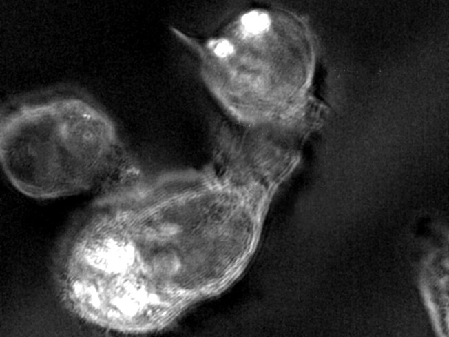
This image, taken by the UT Austin microscope, shows a T-cell (bottom) attacking a tumor cell (top right).
Packed inside every tiny cell, there’s a nucleus, DNA, RNA, rocketing vesicles, walking vesicles, proteins, mitochondria and other biological structures bustling with activity.
And because of the diminutive size and constant motion of these biological structures inside and surrounding a living cell, it remains difficult even for the most advanced microscopes to deliver an image of them.
So, Thomas Milner, professor in the Cockrell School of Engineering at The University of Texas at Austin, and Martin Poenie, associate professor in UT Austin’s College of Natural Sciences, are working to give scientists and engineers a sharper image of this tiny, moving cellular scene.
They are developing and testing a new light-based microscope that they believe is an improved platform for viewing cells, viruses and other biological structures.
Their goal is to develop a microscope that can advance scientific knowledge and help scientists and engineers diagnose diseases and develop new life-saving treatments.
While the microscope is still under development, the researchers have already been able to see viruses emerging from living cells. And they are attempting to image the cytoskeletal structures inside T-lymphocyte cells to gain a better understanding of how they kill tumor cells. Other researchers have been able to image T-cells interacting with tumor cells, but have not yet been able to see inside a T-cell.
The UT Austin microscope can produce images that show structures as small as 25 nanometers (about 3,000 times smaller than the diameter of a human hair) for sustained periods of time, without the use of fluorescent dyes or other contrast agents.
“With our new microscope, you can really see structures that you can’t normally see with current microscopes due to their size,” Poenie said. “For example, you can see individual microtubules that are 20 to 25 nanometers in diameter, and you are seeing these objects without stains or contrast agents going onto the specimen.”
Their new light-based microscope relies on spectroscopy, the use of different colors of light or wavelengths of light to extract information about an object and create an image. Specifically, the microscope takes advantage of polarized light, a type of light wave, to generate contrast with a biological sample and to help deliver an image or picture of a structure that scientists can use.
A major shortcoming of other advanced microscopes is that they rely on fluorescent dyes for contrast, and these fluorescent molecules typically offer only seconds of continuous imaging at a high resolution. The UT Austin microscope, however, offers continuous imaging of a cell for hours without the need for contrast agents. In addition, the polarized light generates sharper images than those that use fluorescent dyes.
“You see all sorts of things in and around the nucleus where things have never been observed. You can see vesicles light up, presumably because of the collagen inside them,” Milner said. “It’s just amazing what you can see.”
Milner and Poenie are still working to refine their microscope and collect data on its capabilities. But they also believe their microscope could be better than existing technologies at producing images of specific structures, such as viruses. To date, scientists have struggled to successfully use fluorescent dyes to tag a virus that can be consistently utilized in the diagnosis of an infection.
In the near future, they hope to develop algorithms to display their images in real time, which would allow scientists and engineers to leverage even greater information about an object than is now possible.
Milner and Poenie have founded a new startup, called Spectropol, to commercialize this technology.