A new microscopy technology developed by researchers from The University of Texas at Austin produced one of the largest and most detailed optical images of complex biological tissue ever created.
They accomplished this feat—without any dyes or labels, revealing the tissue’s natural structure exactly as it is—by creating a computational model combined with a light-based device that overcomes a pesky bottleneck in microscopy. And it does so without using bulky, expensive lenses, opening the door for the technology to eventually move from the lab into the field, where it could be used for global health, education, and public safety applications.
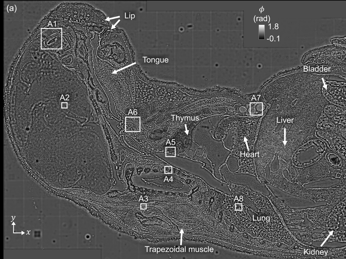
“In microscopy, you typically have to choose between a large field of view or high resolution,” said Sibi Chakravarthy Shanmugavel, first author of the new research published in Optica and a Ph.D. student in the Cockrell School of Engineering’s Department of Electrical and Computer Engineering. “Overcoming that trade-off allows scientists to observe individual cells at high resolution, while simultaneously capturing the larger context of the entire tissue section.”
In the end, the researchers created a 2.5 billion-pixel, or 2.5-gigapixel, tissue image covering an area of 2.7 by 1.7 square centimeters—one of the largest and most detailed images of its kind ever created. This method, which the researchers will continue to build on, is not only simpler and more affordable but also eliminates the need for chemical labels or dyes, making it safer and faster.
To make this happen, researchers placed a thin tissue sample directly on an imaging sensing chip and shone light on it from different angles using an LED array. Advanced computer algorithms then process the data to create clear, detailed images of the tissue. By refining both the image and the illumination angles during reconstruction, the method enables uniform, high-quality imaging across the full sensor area.

“This was a two-dimensional image, and our next phase in this research is 3D computational imaging, further expanding the biomedical capabilities of compact lensless microscopes,” said Shwetadwip Chowdhury, professor of electrical and computer engineering and lead researcher on the project.
Using this on-chip imaging platform, the researchers demonstrated low-cost, high-throughput, label-free imaging across a broad range of biological specimens, spanning multiple tissue and organ types at varying scales. Moving forward, this technology can have strong potential applications in digital pathology, histology, developmental biology, and large-area tissue atlasing.