
Putting Together a Puzzle of Light
Researchers advanced a promising yet challenging light-based method for imaging large, complex tissue samples.
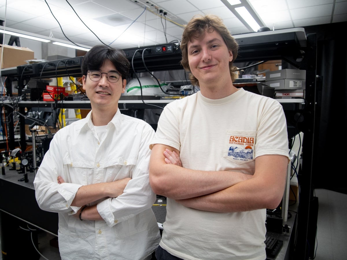
Texas Engineering students Jeongsoo Kim and Peter Wagenaar.
Shine a light on a solid object, and what happens? The focused beam bounces off or through that object, rather than simply passing through to the other side. Pull out a flashlight and give it a try. We’ll wait.
This is the concept of scattering, and it’s a huge challenge for engineers and scientists trying to capture images of complex, multicellular structures within tissue. As light travels through tissue, it immediately bounces through cell membranes, fat droplets and other structures, scrambling an image beyond recognition.
However, the scattered light leaves breadcrumbs that can be reassembled – it’s just difficult to do in practice. Researchers from The University of Texas at Austin have advanced a technology, known as inverse scattering, to do exactly this on two fronts.
First, they established a framework to computationally disentangle scattering effects, enabling robust scatter-corrected imaging in real biological specimens. And they extended this approach to capture how much light a sample absorbs, which is where much of biology’s chemical and functional information lives.
“Now, instead of physically slicing, clearing, or labeling the tissue, we can use computational methods to generate scatter-corrected 3D images from sets of noninvasive scattering measurements,” said Shwetadwip Chowdhury, assistant professor in the Chandra Family Department of Electrical and Computer Engineering and the faculty lead on the studies. “This broadens the range of biological specimens that we can image in their native three-dimensional context. This could be really valuable for studying dynamic processes deep within living tissue, where invasive manipulation can disrupt the very biological processes we want to observe.”
Both studies are steps toward a longer-term goal: microscopes that see millimeters into living tissue rather than tens of microns, using computation instead of dyes or surgery. Nearer-term, the techniques suit organoids and small model organisms, which are used for drug screening, developmental biology and toxicology, where fluorescent labeling is often the limiting factor for watching the same specimen over longer periods.
Better Reconstructions
In a study published in Science Advances, the researchers focused on reconstructing a thick sample’s refractive index, a measure of how much a sample bends and slows light. Reconstructing this gives a fully volumetric and label-free anatomical image of the specimen using its own physical properties.
However, this approach involves solving large, nonlinear optimization problems that are highly sensitive to minor algorithmic choices. The researchers found that small implementation decisions can impact the performance of inverse-scattering reconstruction algorithms in biological samples.
To test this, the team turned to 3D-printed test objects built by collaborators at the Warsaw University of Technology. Each object contains a known cell-shaped target with features as small as 300 nanometers, buried inside a cube of randomly arranged rods designed to scatter light. By printing cubes of 40, 60, 80 and 100 microns, the researchers could dial the difficulty up and down while knowing exactly what the correct answer looked like.
After fine-tuning and validating their methodology on these test objects, the team demonstrated its imaging capabilities on real-world biological samples, including C. elegans, zebrafish embryos, and organoids, which are widely used to study development, disease, and cellular processes. Notably, these samples can be hundreds of microns thick – well beyond the depths at which commercial systems can typically provide high-resolution imaging. In contrast, the team’s method enabled comprehensive, label-free 3D visualization throughout the samples, revealing their internal anatomy in full.
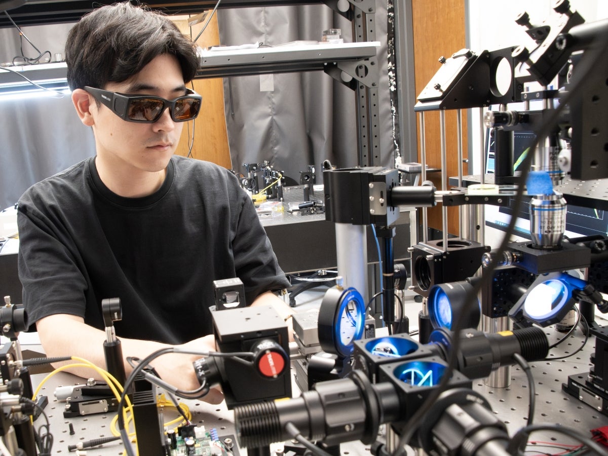
“What’s exciting about this work is that we really pushed the method to see where it could go on challenging, highly scattering samples,” said Jeongsoo Kim, a Ph.D. in Chowdhury’s lab. “We tested and tuned the approach across a large and diverse set of phantoms, and then looked at its performance in actual bio-samples. We found that it worked remarkably well across a broad range of conditions. This gives us a strong foundation to keep pushing the limits of how deep we can image and to see what new biological questions we can tackle with it.”
The project team for this research included faculty members and students across three Texas Engineering Departments as well as colleagues from the Forty Acres and beyond. Others include: Johann K. Eberhart, Elif Sarinay Cenik, Blythe Bolton and Mary E. Swartz of the Department of Molecular Biosciences; Adela Ben-Yakar, Khashayar Moshksayan and Rishika Khanna of the Walker Department of Mechanical Engineering; Sapun Parekh and Mohini Kamra of the Department of Biomedical Engineering; Michał Ziemczonok and Małgorzata Kujawińska of the Warsaw University of Technology; and Karin A. Jorn of Precision One Health Initiative at the University of Georgia.
Shining a Light on Absorption
A second paper, published in Optica, continues on this path. In this research, the team added another dimension to the index: light absorption.
In its original implementation described above, the inverse-scattering framework does not account for absorption, which can lead to errors when reconstructing an absorptive sample. In one experiment, reconstructions of dyed microspheres yielded jagged spikes of impossible refractive index values when absorption was not accounted for.
To fix this problem, the team re-derived the theoretical framework for image reconstruction to explicitly reconstruct both the sample’s 3D refractive index and its 3D absorptivity. They then demonstrate this approach across a range of biological samples, such as algae, plant tissue, and zebrafish embryo.
This is the first demonstration of reconstructing absorptivity alongside refractive index in a thick, heterogeneous sample. This development is particularly important because biological tissues are typically both strongly scattering and absorptive, and absorption can provide information about tissue composition and physiological state that cannot be captured from anatomy alone. By recovering both types of information, the approach further strengthens the impact that inverse scattering can have on scientific imaging.
“Looking ahead, we’re particularly excited about extending this approach toward hyperspectral imaging,” said Peter Wagenaar, a graduate research assistant and one of the lead researchers on the paper. “Because different molecules and tissue constituents have wavelength-dependent absorption signatures, recovering 3D absorption across many wavelengths could provide a molecular fingerprint of what is inside the tissue—not just where structures are, but potentially what they are made of.”
The project team includes Wagenaar and Kim of the Chandra Family of Department of Electrical and Computer Engineering and Swartz and Eberhart of the College of Natural Sciences’ Department of Molecular Biosciences. In addition to funding from the Cockrell School and UT, the project was supported by the National Institutes of Health and the Chan Zuckerberg Initiative.