At the tiniest of scales, it's nearly impossible to capture images of active samples of biological and physical systems, such as living cells, flowing fluids or moving particles. Researchers at The University of Texas at Austin just took an important step in addressing this problem.
They developed an innovative new technique that allows accurate space and time reconstruction of optically scattering samples that undergo simple movements while being imaged.
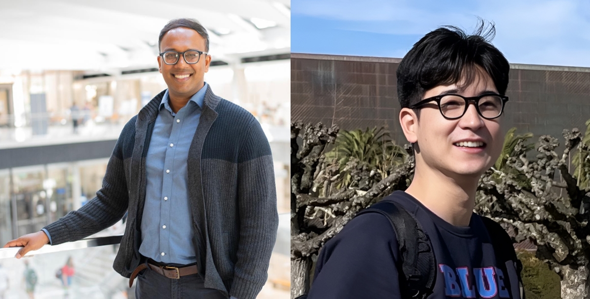
“This is like solving two puzzles at once,” said Shwetadwip Chowdhury, a professor in the Cockrell School of Engineering's Chandra Family Department of Electrical and Computer Engineering who conceived the project. “We’re figuring out what the sample looks like and how it’s moving. By combining these two pieces of information, we can create accurate 3D images even when the sample is in motion.”
The new research was recently published in the journal Optica.
Optical imaging is a powerful tool used in fields ranging from biology to materials science. One advanced method, optical diffraction tomography (ODT), creates 3D images by analyzing how light interacts with a sample. ODT is non-invasive, meaning it doesn’t require dyes or labels, and provides detailed information about the internal structure of samples.
However, traditional ODT assumes that the sample remains perfectly still during imaging. When samples move, the resulting 3D images are often distorted, making studying dynamic systems difficult.
To combat this problem, the researchers developed a joint optimization problem that aims to simultaneously solve multiple, interrelated problems. To do this, they developed physics-based models to simultaneously account for the sample's composition and movement. Unlike some other methods, their approach doesn’t require training on large datasets, making it more adaptable and efficient.
The team demonstrated their technique on various samples that undergo frame-to-frame lateral movement. In one experiment, they imaged microspheres moving in a controlled pattern and successfully reconstructed their 3D structure with high accuracy. In another test, they imaged a more complex sample—a scattering phantom designed to mimic biological cells—and were able to resolve fine details even as the sample moved.
“This is our first step towards studying dynamic processes in multicellular samples, where space and time information is scrambled due to optical scattering," said Jeongsoo Kim, a Ph.D. student in Chowdhury’s lab who spearheaded the project. "Unscrambling this space-time information could lead to better capabilities to image biological processes that occur within their native tissue habitats.”
The researchers emphasized that their method is not only effective but also practical. It builds on existing ODT technology, meaning it could be integrated into current imaging systems without requiring any hardware changes.
While the new method represents a significant advance, the researchers are already considering future challenges. For example, their current technique focuses on samples that move only laterally, but many real-world systems involve more complex motions, such as rotation or non-rigid deformations.


