Magnetic Resonance Imaging (MRI) has been a vital diagnostic tool for decades, aiding in detecting and treating conditions spanning from brain disorders to foot injuries. Despite the benefits, accessibility and affordability continue to pose significant challenges for many.
A new grant from the National Institutes of Health will support the development of novel MRI scans for detecting kidney disease. Led by Adam Bush, assistant professor in the Cockrell School of Engineering’s Department of Biomedical Engineering, and Jon Tamir, assistant professor in the Chandra Family Department of Electrical and Computer Engineering, the five-year, $2.6 million project will focus specifically on Black populations, where kidney disease and its complications are more common.
Why it Matters: Kidney disease is a leading cause of death in the U.S. and affects 1 in 7 adults — 90% of whom are unaware they even have the disease. Hypertension and diabetes are the most common causes of kidney disease, and the CDC estimates that treatment in 2019 cost Americans more than $100 billion.
Glomerular filtration rate (GFR) measures how well kidneys filter waste from blood to create urine, and it is the most common way to assess kidney function. However, GFR tests done through blood samples can be inaccurate, and more elaborate methods are cumbersome, costly, time-consuming and less safe.
Consequently, early stages of kidney disease often go undetected and are rarely studied.
Black Americans are at particular risk. Kidney disease is 15% more common among Black Americans and they are three times more likely to develop end-stage renal disease compared to non-Black Americans. Black Americans also have higher rates of hypertension and diabetes, yet the exact mechanism of kidney disease in this population is understudied.
The Goal: Bush and Tamir aim to develop safe and rapid ways of measuring kidney function using MRI. The technology is widely used to assess the brain but is used in the body to a lesser degree. Key limitations for body MRI applications include physical variation and motion.
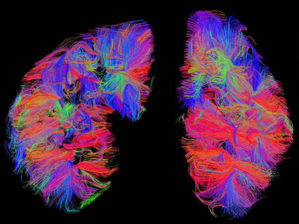
Bush and Tamir aim to solve technical challenges with imaging the kidney, including long scan times and subject movement. The key to their approach is the design of fast and motion-robust sampling combined with advanced biophysical modeling of the kidney.
The researchers believe these methods can create improved indicators for kidney health that are safer, simpler and more precise than current methods for measuring kidney function.
"Imaging the kidneys is challenging, but we've assembled a fantastic team, and we're eager to improve equity and broaden access to historically underserved populations," said Tamir.


